Soft matter optics laboratory keeps a collection of specialized complementary setups devoted to studying the structure, physical properties and dynamics of soft materials. Commercial equipment is continuously supplemented with custom-built instrumentation and newly developed methods.
Location: Room C114
Photon correlation spectroscopy (PCS, DLS)
Responsible person: Alenka Mertelj alenka.mertelj@ijs.si
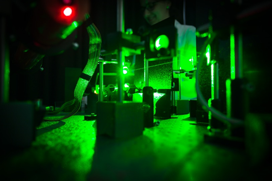
Dynamic light scattering (DLS) from various sample media (solutions, liquid crystal cells, thin surface and free standing films, composite media, etc.) is analysed using an ALV-6010/160 digital correlator that enables measurements in the range of the range of 5ns to ms. Light source can be swithched between a He-Ne laser (632.8 nm) and gren NdYAG (532nm) to minimize absorbance. The normalized intensity autocorrelation function of the scattered light is detected. Different types of scattering geometries and scattering angles in the range 0-140o can be selected. The temperature of the sample can be varied from room temperature up to 150 oC. By combining incident beams from different laser sources the setup can be extended to dual-colour cross-correlation spectroscopy.

Polarizing optical microscopy (POM)
Responsible person: Luka.cmok@ijs.si
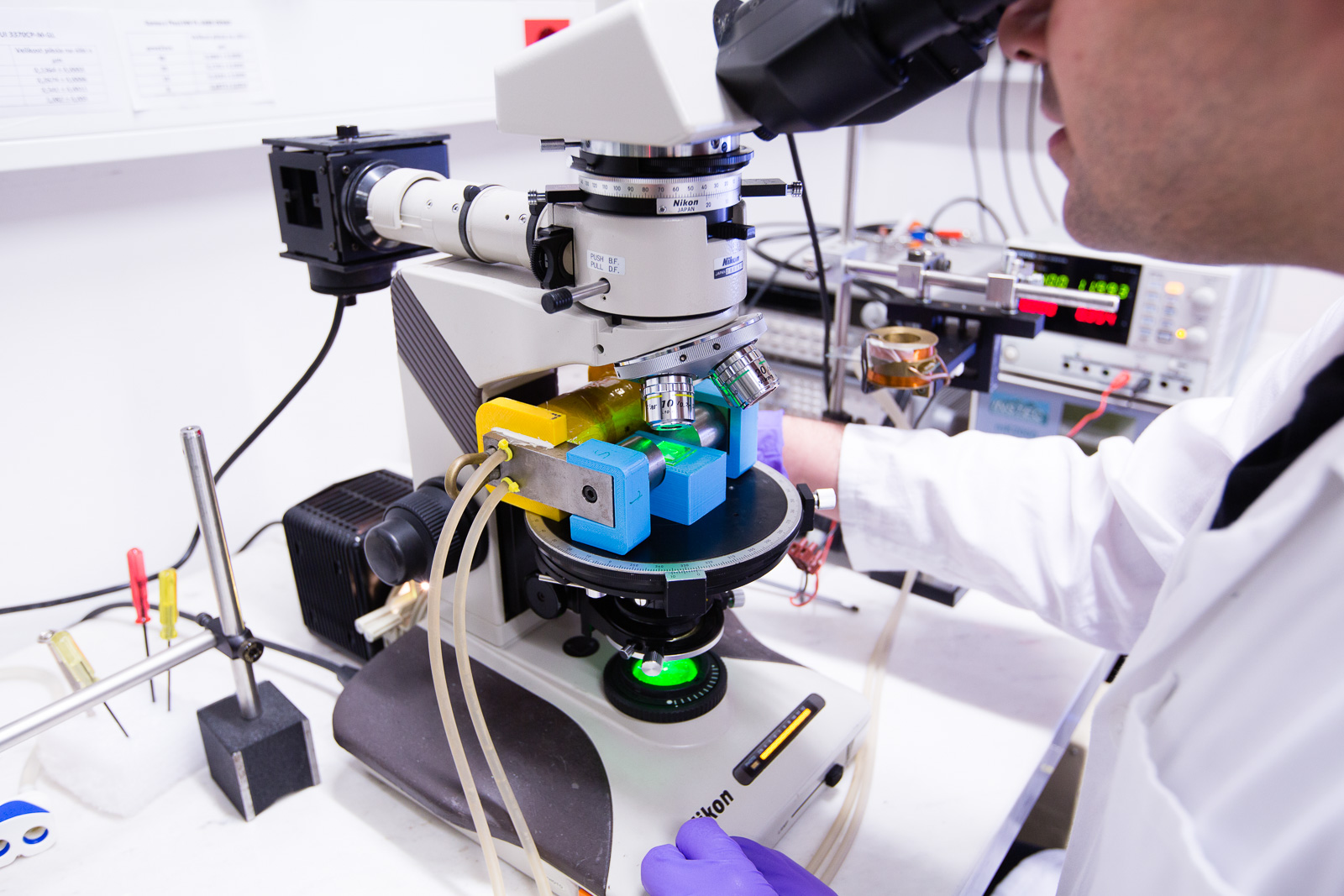
In addition to several custom-made flexible polarizing optical microscopy setups designed to adapt to a variety of systems and targeted investigations, several polarizing microscopes are available including inverted Nikon Eclipse and upright Optiphot-2 POL Nikon. With custom and commercial accessories for temperature control and for investigations under electric and magnetic fields.

Cross-Differential Dynamic Microscopy (c-DDM)
Responsible person: Andrej Petelin andrej.petelin@ijs.si
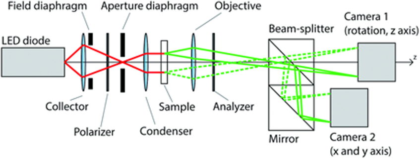
c-DDM setup was developed in our laboratory as extensively described in reference (Arko M, Petelin A. Cross-differential dynamic microscopy. Soft Matter. 2019;15:2791–2797.) by Arko and Petelin. The setup comprises an infinity corrected microscope objective with a 20× magnification, a 20 mm focal length tube lens followed by a beamsplitter, and two identical cameras. Translational and rotational mechanisms ensure precise alignment of their field of view. For illumination, we use the Koehler illumination setup with a nominal wavelength of 565 nm and a bandwidth of 100 nm. The LED is operated in a pulsed regime with a low-duty cycle to prevent sample heating. Cameras are triggered as described in the above reference to acquire two different sets of images with varying times of acquisition. The shortest time delay between two consecutive frames on cameras can be set to 256 μs. The image sequence is then cross Fourier analysed as a function of time delay using an open-source package ‘cddm’ to obtain the normalised image cross-correlation function (Petelin A. Cross-differential dynamic microscopy v. 0.2. Available from: https://zenodo.org/record/3800382#.YEXzkGhKgzU).

Optical second harmonic generation (SHG)
Responsible person: Irena Drevenšek Olenik irena.drevensek@ijs.si & Natan Osterman natan.osterman@ijs.si
Two different SHG setups are currently available in our laboratory. For the first one, the incident radiation are amplified light pulses originating from the Coherent-Mantis/Legend Ti:Sapphire laser system (35 fs long, 2 microjoules per pulse at 800 nm) with OPA/NOPA upgrade(wavelength range from 200nm to 13000nm). Second harmonic radiation generated in the direction of specular reflection is detected by a photon counting system (Andor i-Star) synchronized with the laser amplification unit or a high-performance CMOS camera is used to image large parts of the sample at once. The second setup consists of a custom-built SHG sample-scanning microscope with a Erbium-doped fiber laser (C-Fiber A 780, MenloSystems, 785 nm, 95 fs pulses at a 100 MHz repetition rate) as the laser source. The system additionally allows for SHG interferometric microscopy measurements by insertion of a reference SHG active crystal. The final SHG-M and SHG-I images are acquired with a high-performance CMOS camera or SPAD detector.

Experimental Soft Matter Laboratory at FMF
Responsible person: Natan Osterman natan.osterman@ijs.si
Laboratory dedicated to the investigation of physical properties of soft matter materials providing an extensive additional set of tools and equipment including optical tweezers (one mounted on a fluorescence microscope, the other on a microscope with a magnetic module so the instrument is actually magneto-optical tweezers), fluorescence microscopy (motorized Nikon Ti2-E Widefield Fluorescence Microscope with Hamamatsu Orca Quest camera), polarized microscopy, laser system for microstructuring (soft-lithography and photopatterning of LCs), a cryomicroscope, microfluidics chips fabrication facilities and high-speed camera (Photron Nova S9, 9000 fps at full frame). For additional information please visit: http://tweezers.fmf.uni-lj.si/
